Organ mimics are helping decode complex diseases
On the lab bench, inside a Petri dish nestled between two metal coils, sits an unassuming glob of jelly. But this is no ordinary jelly – it is a squishy gel filled with magnetic particles and cells from the human lung.
Once the power switches on, magnetic currents pass between the two coils, swirling the particles about and stretching the gel out the tiniest bit. When the power’s off, the gel relaxes. Turn the power on and off at a certain frequency, and you’ve got a gel that stretches and relaxes, back and forth, back and forth, almost rhythmically. “It’s trying to mimic a portion of your lung tissue as you breathe,” explains Kaushik Chatterjee, Professor at the Department of Bioengineering (BE) and Department of Materials Engineering, IISc.
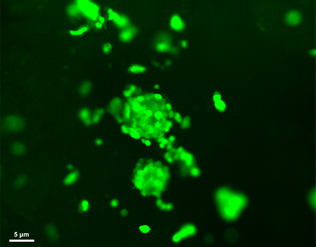
In a science fiction-esque endeavour, researchers like Kaushik are trying to mimic the functioning of human organs in the lab. The “breathing” jelly that his lab is working on is supposed to simulate the action of the human lung. Some researchers are making brain organoids, which are tiny, living blobs of brain cells floating about in a dish. Others are creating solid tumours in the lab by 3D-printing gel-like structures with breast cancer cells, or using patient-derived cells to make gallbladder cancer organoids.
Organ mimics are helping these scientists demystify complex diseases, from cancer and lung fibrosis to TB and schizophrenia
Such organ mimics are helping these scientists demystify complex diseases, from cancer and lung fibrosis to TB and schizophrenia. With labs worldwide increasingly cutting down on the use of animal models, such organ mimics offer an attractive alternative. “From the USFDA to Indian drug regulators, everybody’s pushing hard for alternatives to animal testing,” says Kaushik.
Another reason to switch from animal models to organoids is that they are easier to manipulate. “If I want to modify a gene, or knock it down and go for high-throughput analysis, [it] is not possible using animal models,” says Dwijit GuhaSarkar, Lead Scientist in the Organoid Laboratory at the Tata Translational Research Centre (TTCRC), Kolkata. High-throughput experiments include modifying many genes at once or testing hundreds of drug molecules at a time in a biological system. More importantly, it would be ethically wrong to sacrifice animal models at these scales.
The other issue is that what works in animals doesn’t always work in humans. “So many drugs that seem to be working well in animal models then fail in human trials, and that further slows down discovery,” he adds.
Plus, some diseases like TB, lung fibrosis, gallbladder cancer, and schizophrenia affect humans and animals very differently. Lung fibrosis – a condition in which lungs get filled with dense, fibrous structures that can literally choke people to death – is irreversible in humans, even with medical intervention. But in mice, once the drug that induces fibrosis is removed, the mice’s lungs spontaneously revert to normal. Complex human brain disorders like schizophrenia are pretty much impossible to study in mice models.
“[A] human is not a mouse,” says Rachit Agarwal, Associate Professor in BE. “We need to mimic humans better, whether it’s digitally or through some organoid systems.”
Brain blobs
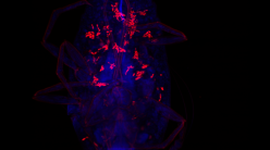
One of the earliest scientists to build a brain organoid was Madeline Lancaster, currently a Group Leader at the MRC Laboratory of Molecular Biology in Cambridge, UK.
In 2013, her team used skin cells from a patient with microcephaly – a condition that leads to shrunken heads and abnormally developed brains in babies – and turned them into stem cells. They then converted the stem cells into neurons and used those to develop cerebral organoids, representing the outermost squiggly part of the human brain called the cerebral cortex. She and her colleagues then compared these patient-derived organoids to organoids made using skin cells from a healthy donor and found that the former could, to some degree, mimic microcephaly. “Amazingly, the [patient-derived] organoids were smaller, and had fewer neurons,” says Madeline, in a 2016 TEDx talk.
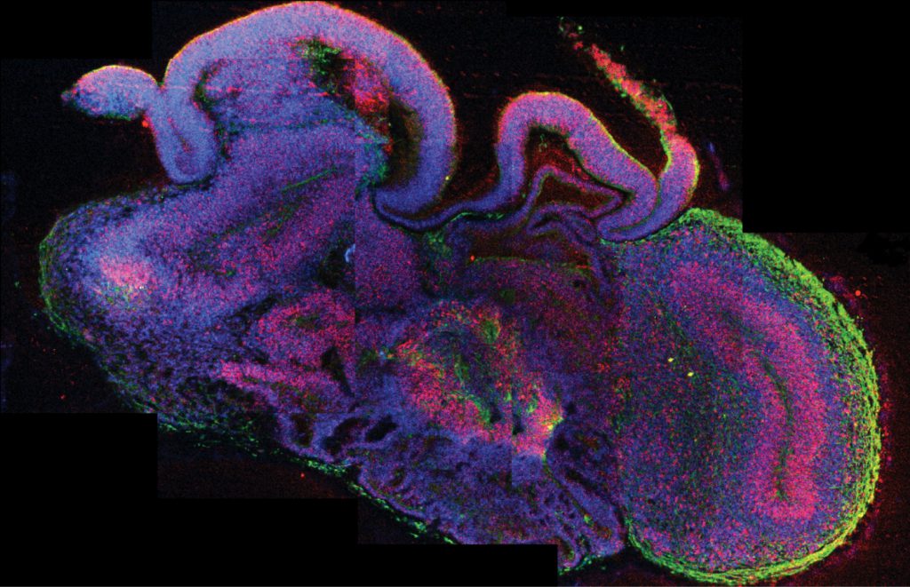
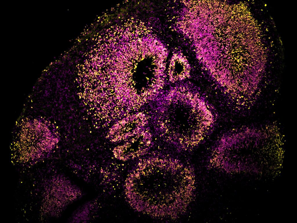
Brain organoids are basically little balls of tissue floating about in a Petri dish or flask filled with a nourishing liquid. But they offer a way for scientists to chip away at one of the most complex organs, “particularly when you want to look at human-specific aspects and also probably capture the genetics of [a] disease,” says Bhavana Muralidharan, Associate Professor at BRIC-inStem, Bengaluru.
A typical starting material for such organoids is human embryonic stem cells (ESCs); extracted from a structure called the inner cell mass in very early stage foetuses, these cells can be turned into almost any other type of cell.
Human induced pluripotent stem cells (iPSCs), like the ones Madeline employed, can also be used. These are adult human skin or blood cells that have been “induced” to become stem cells using specific molecules called Yamanaka factors. iPSCs or ESCs are then treated with a targeted cocktail of chemicals, which gently coax them into becoming neurons. If the chemicals and media conditions are just right, these neurons then self-organise into 3D clumps called organoids.
To dig deeper into how the brain functions, scientists have gone a step further and created assembloids, which are organoids of different brain regions fused together
Researchers can create organoids of specific brain parts as well, like the cerebral cortex, or the entire front part called the forebrain. It all depends on how you nudge the stem cells towards a particular fate – some researchers like Bhavana use a set of molecules that push them into forming smooth cortical spheres. Others don’t use any molecules at all; the media that the cells are submerged in turns them into popcorn-like forebrain organoids. Such organoids can help scientists study how the neurons move around and organise themselves in the developing human cortex.
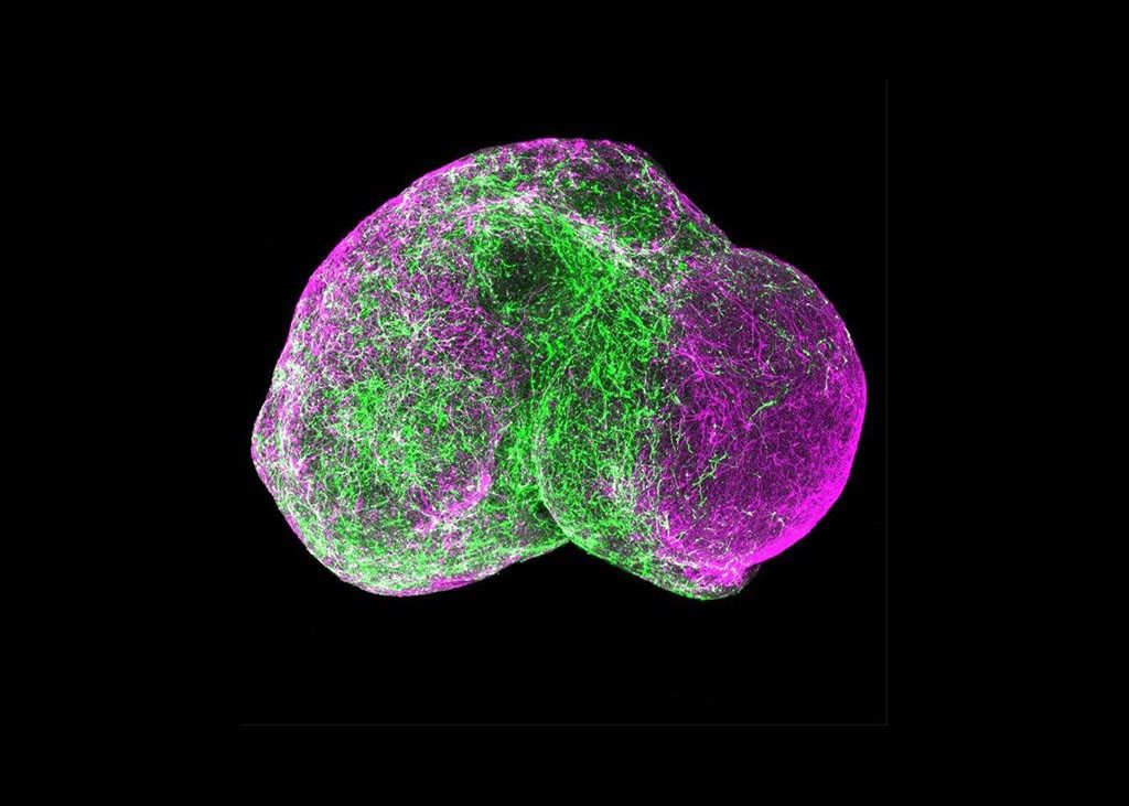
To dig deeper into how the brain functions, scientists have gone a step further and created assembloids, which are organoids of different brain regions fused together. “When you want to look at circuitry, looking at one region of the brain is not enough,” explains Bhavana.
As part of the Accelerator programme for Discovery in Brain disorders using Stem cells (ADBS), now the Center for Brain and Mind, a joint center between the National Centre for Biological Sciences (NCBS) and BRIC-inStem that works with patient families having a history of mental illness, Bhavana has access to iPSCs from people with schizophrenia. She is using these cells to make assembloids and study the neuronal network secreting dopamine in them. Dopamine is a mood-and-movement-regulating chemical, and its release is usually hampered in people with schizophrenia. “You can use these patient samples to come up with a very nice human in vitro disease model,” says Bhavana. Such a model can help explore potential mechanisms underlying any clinical symptoms, she adds. Some dopamine-related schizophrenia symptoms are hallucinations, an inability to experience pleasure (anhedonia), and social withdrawal.
The three brain regions that Bhavana is studying jointly using assembloids are the striatum – a part of the brain that has many dopamine-releasing neurons – the cortex, and the midbrain. Her plan is to first check whether the dopamine neurons are formed correctly in the assembloid. Then, she wants to explore if and how the dopamine circuitry or the connectivity between these three regions is messed up.
Assembloids are the brainchild of Sergiu Pasca, Professor at Stanford University. In 2017, he published a paper showing how his lab fused a cortical organoid with that of the subpallium – a brain region below the cortex that’s involved in foetal development. Once the organoids merged, Sergiu and his team found that inhibitory neurons called interneurons hop across from the subpallium to the cortex and make new connections with the cortical neurons. It almost resembled what happens in the developing foetal brain.
Sergiu’s team also made assembloids using cells from young patients with Timothy Syndrome, a rare genetic disorder that leads to autism, epilepsy, and cardiac dysfunction. When they compared the two assembloids, the interneuron jumps were messed up in the ones derived from patients with the syndrome – the distances were shorter and the interneurons failed to connect with cortical neurons.
In 2020, Sergiu’s team described the fusion of not two, but three organoids – representing a part of the cortex, the spinal cord, and skeletal muscles. When they passed an electrical impulse through the cortical organoid, the muscle portion twitched, indicating that the three blobs had connected.
With such intertwined assembloids, scientists can study complex brain disorders in which the connections between neurons are affected, like autism spectrum disorders, Parkinson’s disease or Alzheimer’s disease, and even obsessive compulsive and bipolar disorders. Once scientists can figure out what circuits may be messed up in these conditions, they can use assembloids to test drugs that might fix the problem, at least at a molecular level. “For instance, we may see that some receptors [are] getting affected. So, can we now look at small molecules to either accentuate or to down-regulate that receptor?” says Bhavana. “That’s where we want to go with this eventually.”
But maintaining organoids in a dish for long is tricky. Unlike the human brain, organoids don’t have arteries filled with fresh oxygenated blood growing inside them. As the blobs get bigger, their cores get cut off from oxygen supply and they start dying out. One solution is to slice up the organoids, open them up to give them some oxygen, and then let them grow back into a sphere, Bhavana says.
“Organoids and assembloids are not full replicas of the human brain, they are not brains in a jar or mini-brains, they are not a stepping stone to some Frankenstein monster,” clarifies Sergiu in a 2022 TED talk. “But using them, we can create avatars of a patient’s brain development.”
“Brainy” organoids
In 2022, scientists at the Australian company Cortical Labs taught neurons in a dish to play the computer game Pong. They wired the neurons to electrodes, which were in turn connected to a computer running the video game. With the right kind of stimulation, the neurons eventually learned to fire electrical signals that could – via the electrodes – manipulate the game’s controls and play it. More recently, the company taught mouse brain organoids how to balance a pole on a virtual moving cart and play the video game Doom (which is more complicated than Pong) – sparking conversations about “organoid intelligence”.
Researchers are also growing brain organoids that last long – Harvard professor Paola Arlotta has organoids in her lab that have been rocking away in the incubator for seven years (the rocking helps better distribute oxygen in the culture medium). These organoids express similar genes as one would find in a kindergartener’s neurons of that age.
The race to build increasingly mature and complex brain organoids has raised concerns over the ethics of growing human brain tissue. Some scientists are worried that the little lumps may eventually become sentient and “suffer” the way humans do, but others counter that “human-like” consciousness – a complex phenomenon that scientists have still not understood – is too remote a possibility in organoids. But issues still remain. “The recognition of possible sentience in honey bees, whose brain size is smaller than some of the more complex organoids in terms of sheer number of cells, grounds the rationale for a continuous monitoring of how size and complexity track with emerging properties,” write a team of researchers and bioethicists in a 2025 policy forum article in Science.
Cancer in a dish
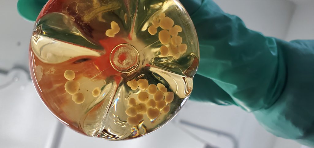
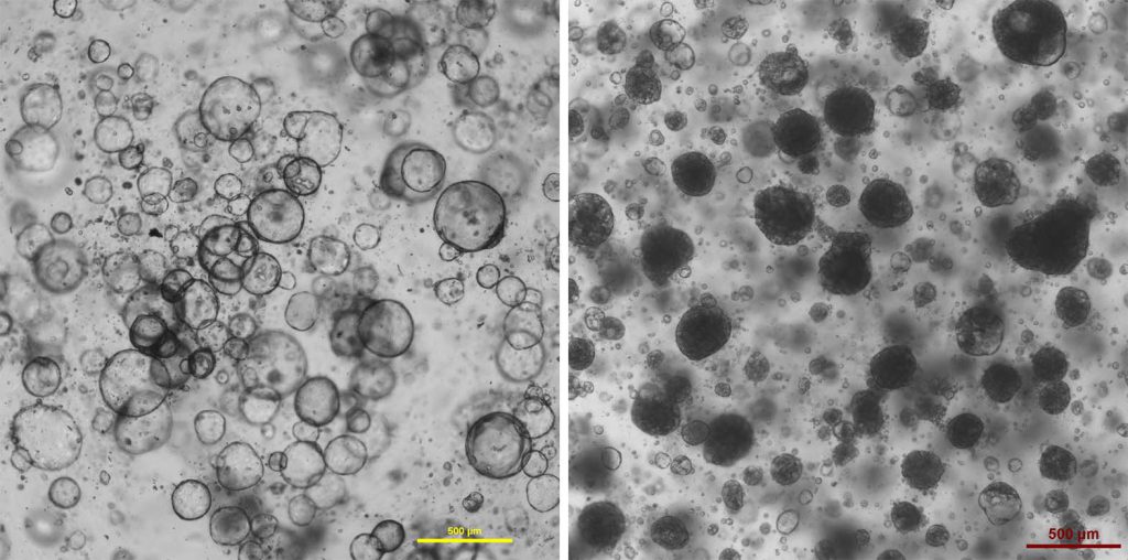
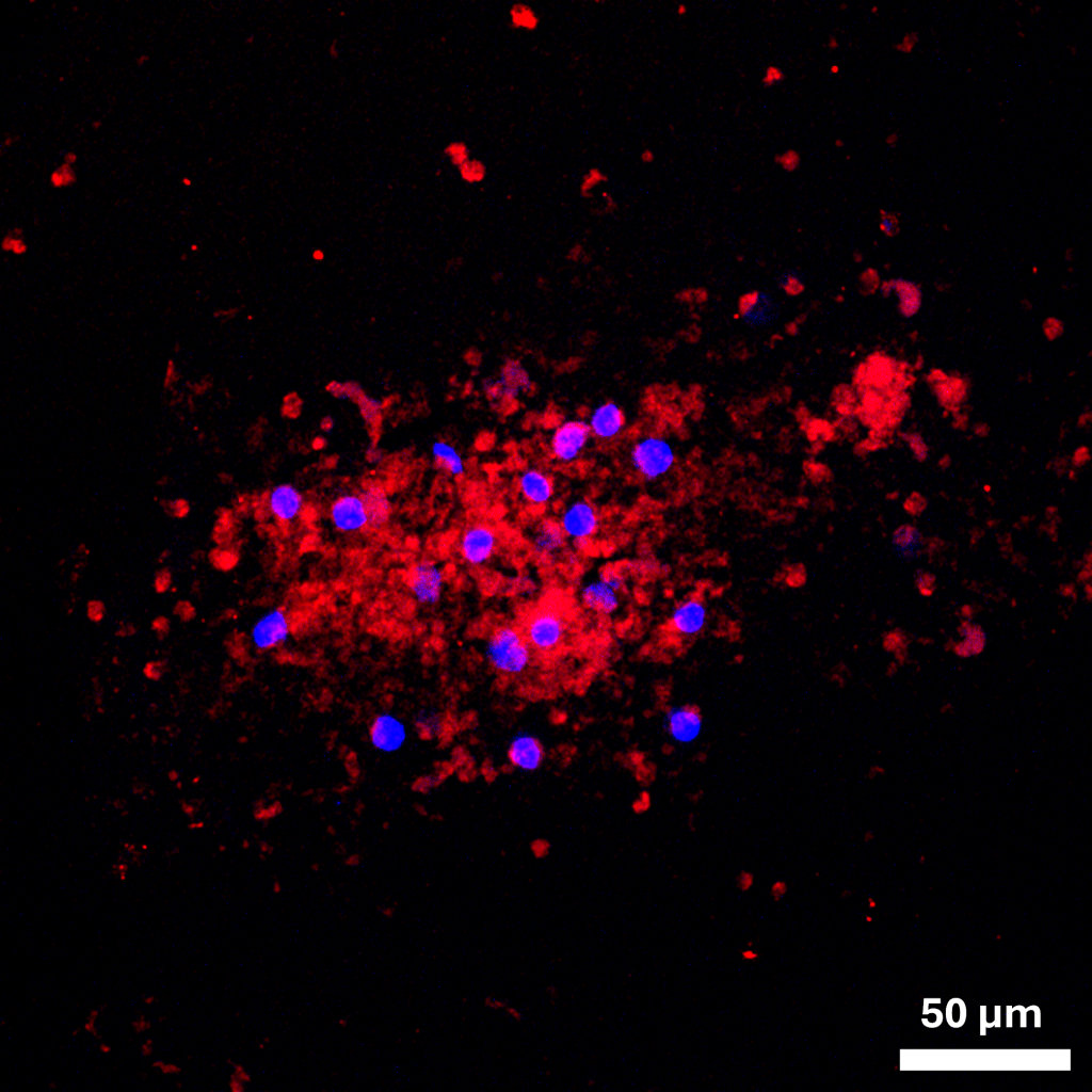
At the bustling Tata Medical Centre (TMC) in Kolkata, within which TTCRC sits, making cancer organoids is a multi-departmental affair. With the help of a dedicated clinical coordinator, Dwijit and his team, called SOLi3d, first keep track of patients undergoing surgery for gallbladder cancer or cancers in neighbouring organs. The surgeon then passes the dissected tissue onto the pathologist, who decides how much of the gallbladder can be spared for research (the first priority is diagnosis). After ensuring that they have the patient and their family’s consent, Dwijit and his team use the cells from the tissue to make gallbladder cancer organoids, which they then store in a biobank. As they are living organoids, researchers can use them whenever they need for experiments – be it DNA or protein analysis or drug testing. “It’s a lot of coordination between the surgery team, the pathology team, the banking team, and the research lab,” Dwijit explains.
Every year, TMC sees 300 patients diagnosed with gallbladder cancer, Dwijit says, and most of them have very poor outcomes. Even though it is a globally rare cancer, it has a high prevalence in the north and northeast of India, particularly around the Himalayan region and the Gangetic belt. Because it is uncommon in the West, gallbladder cancer is somewhat neglected. It is also a silent cancer that can sneak up on people at an advanced stage, making treatment difficult.
Scientists believe that one trigger for gallbladder cancer is chronic inflammation. To investigate this, Dwijit is also growing organoids from patients whose gallbladders are under various stages of inflammation. The idea is to check if and how an inflamed gallbladder eventually ends up becoming cancerous. “Our interest is to study the progression of the disease,” says Dwijit. “So, we are not just growing organoids from cancer patients, we are growing organoids from all different disease states of the gallbladder.”
A 3D organoid version of a tumour is as close as one could get to an actual tumour, making it easier to assess which drugs could treat it
A 3D organoid version of a tumour is as close as one could get to an actual tumour, making it easier to assess which drugs could treat it. The cells are seeded in a dense matrix of proteins, sugars, lipids, and small molecules that resembles the extracellular matrix (ECM) inside the human body – the network of supporting proteins surrounding cells. Just like human tumours grow with the help of chemical cues from their ECM, 3D cancer organoids also respond to molecules from their surrounding matrix as they develop – making them structurally similar to their counterparts in the body.
In addition to gallbladder cancer, Dwijit is interested in using organoids to study breast cancer variants, like triple negative breast cancer (TNBC) – which has no targeted treatment yet – and HER-2 enriched breast cancer – which is extremely expensive to treat. He hopes to develop new, inexpensive drugs and repurpose more affordable, already existing drugs to treat these rare, aggressive cancers.
Using patient-derived cancer organoids can also help us understand how different people respond to the same drugs. “In principle, one can do personalised medicine,” says Kaushik.
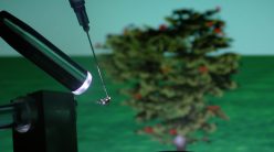
At IISc, Kaushik is also studying breast cancers – he uses gels made up of long chains of repeating molecules called polymers. The cancer cell lines are mixed with these gels and printed onto a 3D scaffold, and the resulting structures can mimic both healthy breast tissue and a breast tumour.
Depending on the ratios of the polymers that they use, and how cross-linked they are, the gel-like material can be stiff or soft. “As a materials lab, we can make various kinds of materials or modify them,” says Kaushik. “We can tune, say, the stiffness of a material to match the mechanical properties of the tissue – for example, if you want a slightly softer gel to mimic the breast tissue versus a slightly stiffer gel to mimic the breast tumour.”
In collaboration with Annapoorni Rangarajan, Professor at the Department of Developmental Biology and Genetics, Kaushik’s team mixed cancer cells from patients with a polymer gel and bioprinted 3D spheres that mimicked a breast tumour. After growing the spheres in the lab and checking if the cells were viable, Kaushik and his team confirmed that standard chemotherapeutic drugs worked well against the tumours.
One can also mix the cells and the gel together to form “bioink”, and then use a 3D bioprinter to squeeze the material out layer by layer, Kaushik says. If you had two types of “ink”, one with cancer epithelial cells (specialised cells lining the tumour surface) and another with immune cells, you could bioprint them as layers to see how the former invades the latter. With new bioprinting technologies, one can even create channels within the material. “So, if I make a tube-like structure, I can perfuse it [with a fluid or blood] and you can think of it as a blood vessel in a breast tumor,” says Kaushik.
The ultimate goal is to replicate the tumour and its environment as closely as possible. “The idea would be to mimic the architecture, the biophysical properties, and the active biomechanical forces,” Kaushik adds.
Lungs in the lab
When TB bacteria attack humans, immune cells are the first to respond. As the bacteria break down the cells’ defences, the immune cells send out alarms, calling out for more of their kind. But the incoming cells only end up becoming more fodder for the invaders. As more and more immune cells flood in to try and quell the disease, they start amassing around parts of the lung. These clumps of immune cells eventually grow to a millimetre or even up to a centimetre in size, becoming what are called TB granulomas. They are what show up as “nodes” in a chest X-ray when someone is tested for TB, says Rachit.
But studying TB in mouse lung models is tricky because the bacterial species that infects humans does not infect other mammals. Primates could work, but studying and maintaining them is expensive and challenging. And, as Rachit says, they are still not human.
Since Rachit and his team came from a materials-based background, they wondered if they could use that to develop an organoid that is more human-relevant. “If we are able to do so, then we can open up several lines of inquiry into understanding and designing therapy against the disease,” Rachit says.
Lung tissue is made up of long chains of proteins and sugars called proteoglycans and biological polymers, one of them being collagen. By carefully selecting which polymers to use, the team created an artificial biomaterial quite similar to the lung in composition, mechanical properties, and softness. “This gives us the initial matrix where we can play [out] this battle between the bacteria and the immune cells,” Rachit explains.
In this artificial lung, Rachit and his team have mimicked the process of how the granuloma forms over several days. So far, their granuloma organoid has grown to about half a millimetre long in size, about five to ten times that of what previous research groups have reported. They also found that some drugs that are prescribed to TB patients work well against the granulomas they grow.
TB bacteria are present in approximately one in four people worldwide, but they remain latent (dormant) in most of them. However, if people with latent TB bacteria end up taking immunosuppressant drugs, then the weakened immune system can end up triggering the bacteria into action. Not all cytokine blockers – drugs that suppress the immune system – re-activate TB bacteria. Rachit and his team also want to use their artificial granuloma to find out which of them do.
“The granuloma is quite a tight structure, so often a lot of drugs don’t penetrate inside and don’t work very well. Because this is an in vitro setup, we can test how deep the drug has been able to go and what the concentration profiles are. Those are the things that we are building this model towards,” says Rachit. “But right now, the major focus has been to characterise and be confidently able to say that this indeed mimics human granuloma and how well it is able to mimic it.”
Granulomas are not the only enemies in the battle to breathe. Sometimes, continuous exposure to pollutants can cause lung tissue to become very thick and stiff. Known as lung fibrosis, this irreversible process can lead to extensive damage, scarring, and breathing difficulties.
In collaboration with Deepak Saini, Professor in the Department of Developmental Biology and Genetics, IISc, Kaushik is keen to use lung mimics to study fibrosis. The idea, he says, would be to mix human lung cell lines with polymeric gels that mimic the lung tissue, add molecules to induce fibrosis, and then use the artificial lung-fibrosis system to test drugs.
Kaushik’s lab has also been studying the effects of air pollution on lung cells. They first bioprinted a gel containing connective tissue cells called fibroblasts. Then, they added lung epithelial cells onto the gel mixture. Using a holder, this gel-cell mixture was then suspended at a slightly elevated level in a Petri dish filled with media. This way, the fibroblasts were submerged inside the media, while the epithelial cells were just at the right level to be at the air-liquid interface, on the air side. To simulate dust, the team sprayed silica particles over the epithelial cells, and found that the cells immediately became irritated. The cells also started producing surfactants to protect the cell surface.
Compared to a classic cell culture setup where one would grow the lung epithelial cells in a flat Petri dish, this setup is much closer to actual human physiology, Kaushik says. His goal is to test what happens to the cells upon prolonged exposure to these silica particles, and to check if fibrosis can be induced, and reversed. “These could be good ways to study lung pathophysiology and also look at drug treatments,” he adds.
Closer to reality
Despite these exciting advances, at this point in time, an organ or tumour-mimic will never truly behave exactly like its counterpart in the human body. “In the body, the brain is connected to every organ, your lungs are connected [and supplying oxygen] to every organ,” Rachit says. “When you isolate an organ like that, there is no crosstalk that is happening with the rest of the organs, which obviously is not the way the human [body] works.” Therefore, some of the results we find using organoids will eventually need to be validated in an animal model or using clinical trials, he adds.
We also still need an animal to understand what a drug does to the body as a whole, Dwijit says, which is why he believes that we cannot completely phase out animal models at this point. “[But] can we reduce the number of animals used in research?” he says. “Because, for certain questions, we probably do not need a living system.” An organoid or organ mimic would be helpful at the preliminary stages of testing whether a drug works or not, before taking confirmed candidates to an animal model or a full-blown clinical trial. “They’re good isolated systems to be able to start to make one’s research more relevant to humans,” Rachit says.
Scientists at Stanford University have already achieved one holy grail – growing liver and heart organoids with a blood vessel (vascular) network inside. Vascularised tissue in the lab brings us closer to better therapeutics, as mimicking blood flow is crucial to take organoids closer to the actual organ. Researchers have also managed to grow blood vessels in skin, lung, gut, and pancreatic islet organoids.
The more realistic organoids are, the more complicated they become
There is, however, a trade-off to making organoids more realistic. The more realistic they are, the more complicated they become, which means that not only are they more challenging to grow, but reproducing them is also tricky. Depending on the application, scientists need to straddle the line between simple but physiologically less relevant organoids and ones that are more complex but definitely more physiologically relevant. While screening multiple drugs to see which one is more effective, for example, a simple organoid may help scientists get reproducible results faster. But a more intricate organ-mimic can help scientists figure out the finer details of how a specific drug might affect an organ.
Which is why the quest to develop the ideal organoid is ongoing. Back in his lab at IISc, Kaushik has found that the lung epithelial cells in his jello grow more when they are dynamically stretched by the magnetic field, as opposed to when they stay still. As the gel “breathes”, its lung cells grow better.
For now, they are only able to keep the experiment going for a few minutes per day for a couple of days, because the gel heats up quickly. “It’s like exercising, you exercise it for a little bit,” says Kaushik. “The cells seem to respond to that, and they do seem to do better.”
Digital doubles
Picture this scene that sounds straight out of a sci-fi movie: A digital hologram of the heart is shimmering above a patient in an operating theatre and doctors are carefully scanning the hologram to determine the best course of action.
Whilst the medical world is not quite swiping at holograms yet, researchers have been able to use computational modelling to recreate the structure and function of some organs, essentially creating a digital duplicate. In a New England Journal of Medicine study from April 2026, researchers worked with 10 patients who had recently suffered a heart attack and had developed a condition that caused their lower heart chambers to beat too fast while pumping blood. Using MRI scans, the researchers digitally recreated 3D versions of the patients’ hearts and then used computer simulations to model how electrical signals will zip through the heart’s muscles. This way, before surgery, they could precisely predict how to operate on and remove the muscles generating the errant electrical activity, which would help correct their heartbeat. This helped reduce the surgery time from three hours to roughly 30 minutes.
Scientists at the University of Liverpool in the UK have also digitally recreated the retina for in-silico drug trials. Others at Wayne State University in Detroit, USA, have modelled the placenta to study issues that could arise during pregnancy. The ultimate goal is to make a whole-body “digital twin”, “a personal guinea pig for testing out medicines”, writes Jessica Hamzelou in an MIT Technology Review feature. Predictably, the prospect of human digital doubles has raised concerns over patient autonomy and who will own such highly personalised data.
(Edited by Ranjini Raghunath)