On research, failure and learning to work with the mess
The experiment, at least on paper, was simple: watch what happens to the physical forces between cells as a healthy tissue starts becoming cancerous. Push, pull, tension mapped onto something as messy as a living tissue.
I remember feeling all tingly when I came up with this idea a couple of years ago, while finalising my PhD research topic – the excitement of stepping outside the usual route that cancer was investigated. Most cancer research I had seen so far was at the level of genes, molecules, and signalling pathways. But here, the question felt tangible. I could actually watch cells pushing against each other, holding shape, and reorganising as cancer takes over in a healthy tissue.
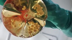
And the way I planned to study it made it feel even more real. Instead of growing cells as a flat layer in a flask or dish, I would grow them in a gel that allowed them to organise in all three dimensions as in an actual breast tissue. I was drawn to breast epithelial systems because of their organised architecture, neat layers, and clear boundaries that made the tissue appear almost geometric under a microscope. Over days, the cells assembled themselves into small, spherical structures called acini, resembling the tiny milk ducts where most breast cancers are known to begin.
If the experiments worked, I would be watching the earliest moments of cancer taking shape and maybe even publish a groundbreaking paper. But a few months into the project, things started falling apart.
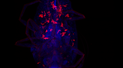
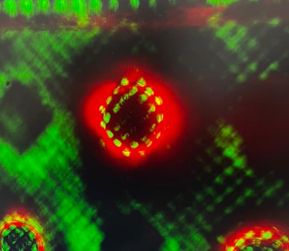
I was in the lab one evening, staring at the screen for so long that the fluorescent green cell outlines blurred into each other. The acini were supposed to have formed neat, hollow spheres with a single organised shell of cells wrapped cleanly around an empty centre. Instead, the images looked cloudy and indistinct, flooded with green haze and fuzzy light yet again! I kept adjusting the microscope’s focus, hoping the blur would suddenly resolve into something meaningful. But … nothing.
The lab was unusually quiet that evening. The hum of the incubator, the faint whirr of a centrifuge in the background, the harsh white light reflecting off stainless steel surfaces. Everything around me felt steady, working exactly as it should.
Except this.
I sat there for a while longer, willing the image to make sense. But I could not ignore the heavy sense of dread creeping into my stomach. Had I just spent the last six months building an experiment that simply wasn’t going to work?
Nothing looks the way it should
Doing a PhD was never part of my career plan. After my Master’s in biotechnology from St Xavier’s College in Kolkata, I had imagined a more straightforward path for myself in industrial R&D – something structured and predictable, with clearer timelines and outcomes. But when my father was diagnosed with cancer, I found myself becoming increasingly drawn to questions around the disease. My interest eventually took shape through understanding how cells physically interact and reorganise as healthy tissue begins to turn cancerous.
Eventually, I joined the Department of Bioengineering at IISc for my PhD, and when my thesis idea of working on the mechanical aspects of cancer was finalised, I was thrilled.
That is, until I hit that snag a few months in, and I suddenly had to rethink my entire strategy. What made it worse was the timing. My comprehensive exam was approaching, and I still had no concrete data to show. For months, I had been building and imaging the acini system, but I still had not figured out how to see the structures clearly enough to extract anything meaningful. Every week that passed made me feel more behind. The project that had once felt exciting now felt unstable. My entire PhD was now resting on an experimental system I still could not properly access.
Maybe I didn’t need a three-dimensional model after all, I started telling myself. If there are forces acting within tissues, they would also exist in simpler cellular systems, right?
So, I took a step back. Moving to the monolayer felt, at first, like stepping away from the biology. The three-dimensional system had always felt closer to the real thing. A monolayer was different. The cells spread themselves flat across the bottom of a dish, more like a thin sheet than the native architecture of biological tissues. It was simpler to image, easier to manipulate, but also further away from the complexity of a real organ. And somewhere in my head, I had started equating complexity with importance.
I spoke to my PI about this, half expecting resistance, but she approached it far more pragmatically than I did and told me that if this was the system that could give us something measurable and reproducible, then it was worth following. That clarity felt reassuring. While a part of me felt like I was conceding, I was also relieved.
The pivot itself took a few weeks. I had to abandon many of the routines I had spent months optimising for the acini experiments and return to much simpler setups. Different dishes, different imaging conditions, different ways of preparing the cells. Not to mention the added stress I felt – for the next several weeks, my days became less about chasing the original idea and more about rebuilding the experiment from scratch. But slowly, as the first usable images started coming in, curiosity began replacing that discomfort.
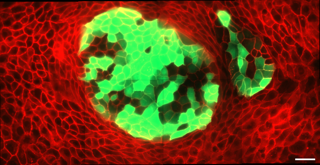
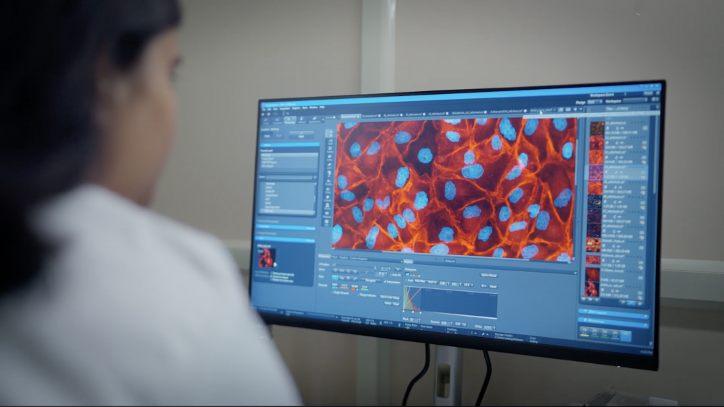
I grew two populations of cells together in the same dish – one healthy and one carrying cancer-associated mutations which were dyed fluorescent green. Over time, the two populations expanded until they met and formed a boundary. During the acini experiments, I would often spend hours adjusting the focus through thick layers of tissue, fighting blur and fluorescent haze just to see what was happening. In the monolayer system, the cells sat exposed in a single plane, their interactions unfolding directly under the microscope. The cell boundaries were sharper. Movement was easier to follow.
The first images from the monolayer didn’t look remarkable. The cells were arranged in familiar patterns, interfaces forming where the different populations met. However, I quickly found myself lingering at these interfaces between the healthy and cancerous cell populations.
Something strange was happening.
The cell boundaries weren’t fixed but were moving, changing, almost as though they were being negotiated. I breathed a small sigh of relief. Something more dynamic than I had expected was happening. I zoomed in and followed the same regions again, frame by frame.
As excited as I was, it took me a while to trust the validity of my observations. Was it noise? It often is. Random fluctuations in the imaging, small inconsistencies from one experiment to another, things that look like patterns until they disappear on repetition.
But the movements didn’t go away. Across different fields of view, across repeat experiments, the same behaviour emerged. The boundary wasn’t passive, it held shape in some places, gave way in others, and reorganised itself.
This was not the assumption I had started with. I had thought that the cancerous cells would simply grow faster and overtake the healthy cells because of their mutations or growth advantages. I wasn’t expecting the interface itself to behave dynamically or mechanically, almost like a physical boundary under tension.
Instead of cancer cells simply overpowering healthy cells because they grew faster, the boundary between the two behaved like an active mechanical interface that influenced whether the cancer cells advanced or were restrained.
The system, in its simpler form, was revealing something.
Finding a new avenue
Something acting at the boundary between healthy and mutated cells played a role in how the cancerous cells behaved. I started with this initial hypothesis and began to test it more deliberately.
I tried to break down the boundary between the two populations – healthy cells and cancerous cells – and watched how the cells reorganised afterwards. Sometimes, the cancerous cells would begin pushing forward collectively, slowly displacing the healthy cells. At other times, the boundary would hold its shape more rigidly, resisting that movement altogether. In some regions, the interface straightened tightly, while in others it loosened and buckled slightly before settling again.
I kept going back, almost expecting them to disappear if I looked closely enough. A different batch of cells, a slightly altered condition, a repeat on another day. Weirdly, each time, a small part of me hoped it would disappear – so I could go back to the original plan, to something that met my initial expectations.
But they didn’t go away. The cells seemed to behave according to some underlying physical rule. I didn’t have the language for it at first. Only later, after many more late evenings spent staring at the same regions of the same images, could I begin to describe it. It was not just movement or rearrangement. It was tension.
Interfacial tension, to be more specific. The forces acting at the boundary between healthy and cancerous cells weren’t just shaping how the interface looked; they seemed to determine what happened next – whether the cancerous cells pushed forward and colonised the tissue, or whether the normal cells held their ground.
And all of it was happening right in front of my eyes on the computer screen. While the idea of physical forces in tissues is not new, what I was seeing suggested something more urgent – that even at the earliest stages, the interaction between healthy and cancerous cells could be shaped by simple physical forces at their boundary. Once I saw it that way, everything else began to align. It was strangely satisfying to observe something so interesting emerge organically from a system I had initially dismissed as too simple.
The experiment hadn’t worked the way I wanted it to. But it had worked. By approaching the original question from another direction, it revealed something more fundamental that might have remained hidden in a more complex system. What had felt like limitations at the beginning – the inability to perturb the tissue and the lack of resolution – had shaped the discovery itself.
Ironically, around the same time, the original acini system, which I hadn’t (rather couldn’t) fully abandon, had also started behaving better. I had been trying to improve the imaging pipeline, and the three-dimensional cultures eventually became far more reproducible. But by then, the monolayer experiments had already opened up an unexpected avenue that I could not stop chasing. The results felt too interesting to abandon midway, so I decided to follow that story first.
Making sense of what I found
When I finally had a complete set of meaningful results, I showed them to my PI. She went through them quietly and said something along the lines of, “There’s something interesting happening here. We should follow it carefully.” That was all. Which meant that it would still be a while before any of this could be woven into something I could call a concrete discovery. The next few months were … different.
There was a different kind of urgency now. Instead of trying to make something happen, I was now desperately trying to understand what was happening. I made small variations while repeating the same experiments, used careful controls, and spent long hours on analysis that didn’t feel as frustrating as before.
Finally, I laid out all the data I had collected in order. I loosely placed the images next to each other. Microscope snapshots of two groups of cells meeting their boundaries shifting over time. I drew arrows along the interfaces, tracing where one population pushed into the other, where the boundary straightened, where it buckled.
Notes scribbled in the margins: repeat? noise? pattern? A story was forming, not in words yet, but in images. Figures were revised repeatedly, often during quick discussions with lab mates or long evenings with my PI pointing out inconsistencies I had missed.
The next challenge was writing all of this down into a paper. This phase demanded a clarity that the experimental process never had. The messy progression, the failed attempts, the shifts in direction, the long stretches of uncertainty, all had to be distilled into something that looked intentional.
I found myself thinking more about why I had started this research in the first place. When my father was going through his own experience with cancer, it was an immediate and lived experience. Appointments, treatments, small routines built around managing something that didn’t have the neat outlines of a research question.
I had entered the PhD assuming that enough effort and persistence would eventually lead to something concrete – something directly connected to helping patients. This was different.
What I had found was more fundamental. A way of thinking about how cancer cells organise and interact with healthy cells. But it wasn’t a solution. Was it enough? For a while, I was caught between the satisfaction of finally having something that worked and the uncertainty of what it meant beyond the experiment itself.
Over the next few months, that tension softened over mundane moments – while explaining the results to lab mates, while sitting with the same set of images for hours, zooming in and out. Even while walking back from the lab after a long day, I kept replaying the same images in my head.
The big picture
The paper itself took shape slowly through months of writing and rewriting, revisions and addressing comments from publication reviewers who weren’t always convinced.
Eventually, after three whole months of back-and-forth with the reviewers, revisions, more experiments to answer unanticipated questions, and countless moments of fear and doubt, my paper was accepted. After two and a half years. I was alone when I got the email. I kept rereading the first line to be sure I hadn’t misread it.
For most of its life, the experiment was uncertain. It resisted, stalled, shifted direction without warning. It asked for patience in ways I hadn’t anticipated and gave answers only when I was willing to change my approach.
The version that exists now, the one that fits into a paper, that can be explained in a few figures and a few paragraphs, is only the final form. But the life of the experiment – its hesitations, its refusals, the way it changed shape under constraint – remains mostly hidden. Yet, that is where most of the work really happened.
Looking back now, I think my most important accomplishment was not the paper or a result but being able to sit with uncertainty long enough to eventually figure something out.
Now, after publishing the monolayer work, I find myself returning to the acini system again for the next phase of my PhD, only this time with a much clearer idea of what I am looking for.
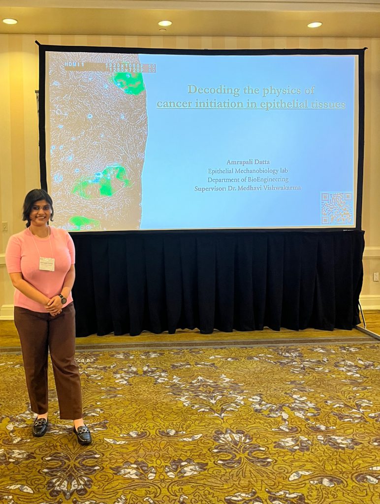
Amrapali Datta is a fifth year PhD student at the Department of Bioengineering, IISc, and a science writing intern at the Office of Communications
(Edited by Abinaya Kalyanasundaram)